Anatomy |
||||||||||||||||||||


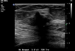
A
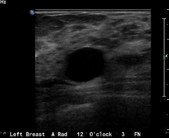
B
Breast ultrasound (US) is the correct next study to order. Image A shows the ultrasound on the patient shown in the last case. Image B is the example of a cyst on ultrasound. How do these two images differ? Think about margins, shape, and what is happening DEEP to the actual lesion on the image.
Using the clock face can be a bit confusing in the breast, since the RIGHT breast is different from the LEFT. If you are facing the patient, the RIGHT 3:00 position will be in the inner breast, while the LEFT 3:00 position will be in the outer breast.