ICM-II: ABDOMEN, more cases |
||
 |
||
 |
|||||||||||
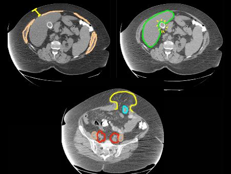
TYPE-CT scan (cortical bone is white), axial imaging plane, soft tissue window (fat and muscle are very different shades of grey, easy to tell apart).
TERMINOLOGY- high density in region of gallbladder (yellow on top right image, this was a single large gallstone), also large ventral hernia (yellow outline on bottom image) containing a small bowel loop. the liver is outlined in green.
TECHNICAL- No IV contrast (vessels and muscles are the same shade of grey), some oral contrast in a few bowel loops but not in all of them (may not have been administered ideally, which would involve slow ingestion over several hours).
Patient body habitus shows much more fat than muscle (top left image) suggesting obesity and perhaps deconditioning.