Anatomy Yr-1 |
||||||||||||||||
25 year old male with left hip pain |
||||||||||||||||
 |
||||
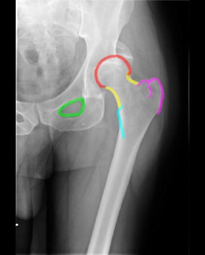
On this AP radiograph of the femur, the femoral head is outlined in red, the femoral neck yellow, the greater trochanter purple (where the gluteal muscles attach), the lesser trochanter aqua (where the iliopsoas muscle attaches), and the obturator foramen green. A general radiographic principle is that for most bones, we like to obtain two images at 90 degrees from each other (orthogonal), like an AP and a lateral view. This gives a better idea of the structure in three dimensions. What is difficult about doing a lateral radiologic view of the hip joint? |
||||