Anatomy Yr-1 |
||||||||||||||
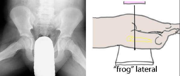
If you just shoot the x-ray beam from the side, both hips will be on top of each other and hard to figure out. There are two other ways to get views of the femur from different angles. The first is called a 'frog' lateral, shown in Images A and B. The femur is abducted to the side, which rotates the bone by about 90 degrees. The white area overlying the middle of Image A is a lead gonad shield to decrease radiation of the testes. |
||||
 |
||||
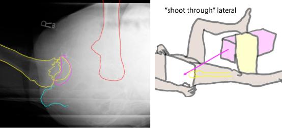
In a patient like ours with a severe hip injury, they will not be able to move their femur into a frog position. The alternative view is called a 'shoot through' lateral, shown in Images C and D. The affected femur is not moved, but the opposite leg is flexed and raised to get it out of the image. The left femur in Image C is outlined in red (flexed and moved away from the affected side), the right femur in yellow, the right acetabulum in purple and the right ischial tuberosity in blue. These images are hard to interpret but may be the only view that can show a severe injury. |
||||
 |
||||
D
C
A
B