Anatomy Yr-1 |
||||||||||||||
 |
||||||
 |
||||||
 |
||||||
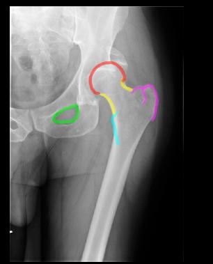
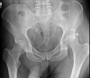
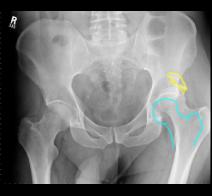
On Image A, as on many types of radiologic studies, it is best to compare right to left for symmetry. If you look at the right hip joint, the space between femur and acetabulum is clearly seen and is a thin lucent line, because cartilage is much less dense than bone, similar to other soft tissues. As shown on Image C, the left femur is not lined up correctly with the acetabulum, but is diplaced upwards. The expected normal location of the left femur is shown in aqua, and there is a fragment of bone superiorly shown in yellow. Image B is a femur x-ray of the same patient, with anatomic structures outlined for you to identify. The femur radiograph shows more of the shaft (diaphysis) of the bone than the pelvis radiograph. |
||
C
B
A
CASE 2