Anatomy Yr-1 |
||||||||||||||||
55 year old male with right scapular lump and pain |
||||||||||||||||
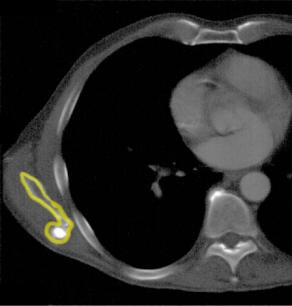
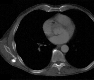
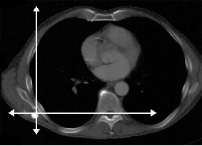
CT is a great way to separate out structures that are overlapping on a radiograph, particularly for imaging bony abnormalities. The lower scapula and the abnormal bump sticking off posteriorly are outlined in yellow on Image B. For the chest radiograph, the scapular position is different than for a CT, where the arms are lifted overhead. In Image C, the scapula has been moved to a position more like that of a chest radiograph and you can see how the bony bump would be seen over the lung on a PA view and over the spine on a lateral view. This is a benign growth called an osteochondroma, and can be removed if it is painful. |
||


A
B
C