|
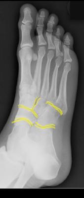
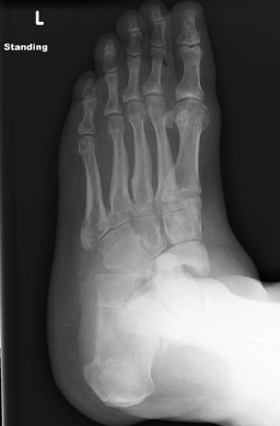
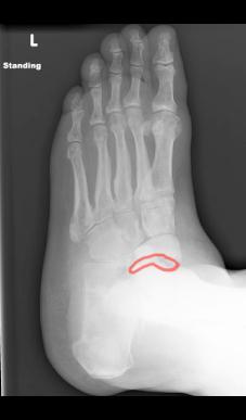
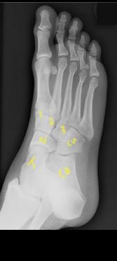
Images A and B are the oblique views of our patient and one of the joints appears too wide (outlined in red on Image B). Compare to the uniform thin joint spaces seen on normal comparison image C and outlined in yellow on Image D. The foot bones are indicated on Image C (1,2,3 cuneiforms, Cu=cuboid, N=navicular, T=talus, Ca=calcaneus). Widening of a joint may indicate disruption of the ligaments that hold it together. The ligaments in the foot are usually very strong and it takes a lot of force to disrupt them. Can you think of a reason that a patient with diabetes might not be aware of repeated damage to their foot?
|
|
|


