|
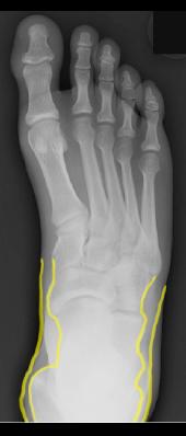
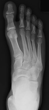
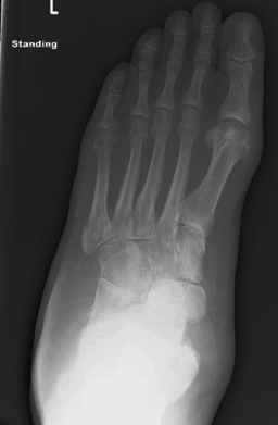
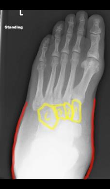
Image B shows soft tissue swelling (outlined in red), compared to normal soft tissues on Image D (outlined in yellow). Normally there is only a thin layer of skin and subcutaneous fat overlying the region of the proximal foot bones and ankle region, but in our patient there is much more soft tissue. On Image B, the cuboid (C) and three cuneiform bones ( 1, 2 and 3) are outlined. If you compare the density of these bones on Image A to the same bones on Image C, you will notice that our patient's bones look 'washed out' or not as dense as normal. This can happen in cases of infection or when patients are not ambulatory (not walking) and the bones begin to waste away from lack of use.
|
|
|


