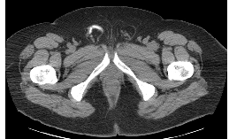
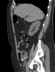
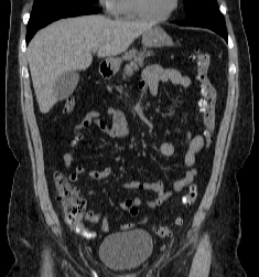
TYPE-CT scan (cortical bone is white), initial images shown in axial plane, soft tissue windows (coronal and sagittal images are shown here.
TERMINOLOGY-a tortuous tubular high density structure is present in the right inguinal canal (looks like a worm)-this is the patient's appendix which is within an inguinal hernia. Patient body habitus looks fairly normal, with just a bit more fat than muscle along the body walls.
TECHNICAL- no IV contrast (vessels and muscles are the same shade of grey), oral contrast (the appendix would be much harder to identify if it was not filled with oral contrast, making it appear very white).


