ICM-II: CHEST, case 4 |
||
 |
|||||||||||
Nuclear medicine studies offer very powerful ways to demonstrate and quantify many different normal and abnormal processes in the body. These images are a 'dynamic' study of ventilation, showing how the radioactive gas is eliminated from the lungs over time. This can be used to show areas of air trapping, as might be seen in emphysema or chronic obstructive pulmonary disease (COPD).
Terminology for nuclear medicine images is different from radiographs or CT scans. If images are displayed with a dark background (as these are), then white areas are areas of increased UPTAKE (also called 'hot' spots), and dark areas show decreased UPTAKE ('cold' spots).
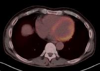
A very useful type of imaging can be done with nuclear medicine and CT at the same time, called PET/CT. The tracer for PET is taken up by many types of tumors as well as some normal tissues, such as myocardium and brain. A normal chest PET/CT image is shown below.


In this fused image, the nuclear medicine information is shown in color, aligned on top of the CT image data. The hot areas are in red/orange. The uptake in the left ventricular wall is normal. Any sites of tumor would show up as focal red hot spots.