ICM-II: CHEST, case 4 |
||
 |
||
 |
||
 |
||
 |
|||||||||||
This is an example of a nuclear medicine study called a ventilation-perfusion study (or VQ scan, for short). Nuclear medicine studies involve introduction of radioactive materials into the body through various routes, followed by placement of a large detector close to a part of the body to determine where the radioactivity is coming from. Many different 'tracers' or radiopharmaceuticals exist to image different organs and processes in the body. Other tracers can show bones, heart, or liver, as well as tumors or infection.
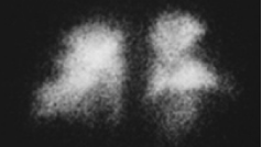
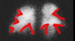
This is the perfusion part of the abnormal study. A tracer is injected into an arm vein. The tracer is designed to be just large enough to get caught in the first capillary bed it encounters, in the lungs.
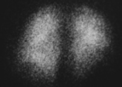
This is the ventilation part of the study. A tracer is inhaled, to show where the ventilated parts of the lungs are. The ventilation image should look very similar to the perfusion image in a normal case.
In this case, there are many peripheral defects in the perfusion study, indicating pulmonary emboli. The two images shown were obtained from a posterior view, to show the deepest and lowest parts of the lower lobes of the lung. The detector could also be placed adjacent to either side of the chest or the front of the chest to obtain other views.