![]() |
![]() |
![]() |
![]() |
![]() |
||||||
![]() |
![]() |
![]() |
![]() |
![]() |
![]() |
![]() |
![]() |
![]() |
![]() |
![]() |
ICM-II: CHEST, case 4 |
||
![]() |
![]() |
![]() |
![]() |
||||
![]() |
||||
![]() |
![]() |
![]() |
![]() |
![]() |
![]() |
||
![]() |
![]() |
![]() |
![]() |
|||||||||||
![]() |
|||||||||||
![]() |
|||||||||||
![]() |
|||||||||||
![]() |
|||||||||||
![]() |
|||||||||||
![]() |
![]() |
![]() |
![]() |
![]() |
![]() |
![]() |
![]() |
![]() |
![]() |
![]() |
![]() |
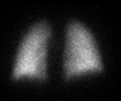
These are two examples of a different type of imaging that can be done to diagnose PE. Which is the normal study, and what are these types of images called?
This is a second part of the study done in our patient. How was this study done? What does it contribute to the diagnosis?