ICM-II: CHEST, case 4 |
||
 |
|||||||||||
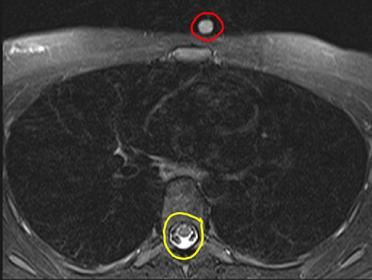
The bones on this image are not as easy to see as on a typical CT scan, and each bone has a dark rim around it. This is an MR image in the axial plane. MR images can either have bright fat or darker fat (as in this image) due to 'fat suppression', which lowers the signal from fat. Because the fat and muscle are similar in signal, it is hard to tell how much of either is present in this patient. MR can also be used to detect PE, but it is more expensive and less available than CT, and is not easy to do on unstable patients who require monitors.
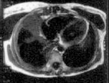
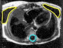
The images to the right are from a different patient, who has saline breast implants. The fluid in the implants as well as the CSF are both low signal, so this is a T1-weighted image. There is no fat saturation, so the fat is high signal.

The object outlined in red is a special MR marker to show where the patient had pain. The study is normal. The marker for MR cannot be made of metal, like CT markers, since that would produce artifact on the image. The markers often contain fatty material, like vitamin E in a capsule, which will give higher signal on MR.
To determine whether this is T1 or T2-weighted imaging, you need to find some water or watery fluid. The cerebrospinal fluid if often a good place to look (outlined in yellow), showing high signal and indicating that this is a T2-weighted sequence.

