Anatomy Yr-1 |
||||||||||||||


One more case with a renal abnormality...
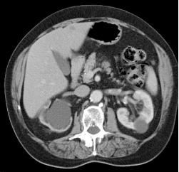
Patient D has a very dilated right renal pelvis and calyces with very little remaining right renal cortex. This is due to obstruction, which caused the kidney to shrivel up (atrophy). The cause of this patient's distal ureter obstruction was accidental clipping during a hysterectomy.
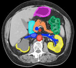
Labels for Images E and F: purple-stomach green-colon yellow-kidneys, orange-pancreas aqua-duodenum dk blue-renal veins, SMV red-aorta, SMA
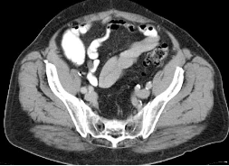
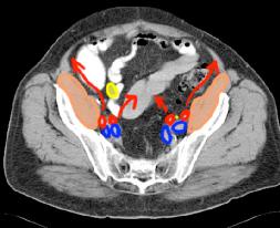
In the pelvis, the right ureter is dilated while the left is normal (small and hard to see). The psoas muscles and internal/external iliac arteries and veins are shown on Images G and H.
Patient D
E
F
G
H