Tufts Anatomy Yr-1 |
||||||||||
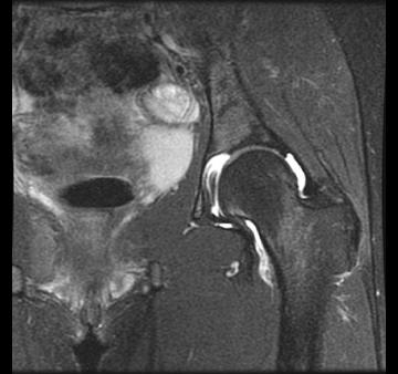
MR or magnetic resonance imaging is another modality that is often used in musculoskeletal imaging. It images entirely different tissue features, not DENSITY as is used in CT. Cortical bone is very black on MR, the opposite of CT, where cortical bone appears white. Fluid on MR can appear dark or bright (as in these images) based on how the scan is performed. |
||||
 |
||||
This is a coronal MR image on a different patient, showing fluid around the hip joint. MR is best for many soft tissue structures, as it images based on various chemical/magnetic properties of tissues, and is often good at detecting fluid, either in a joint space (like in this case), intestine, bladder or in other regions such as sites of infection or trauma. |
||
B
A
CASE 1 followup