Tufts Anatomy Yr-1 |
||||||||||
CT data can also be reformatted to produce rotatable 3D images, such as those shown in Image B. These can be very useful for surgical planning as they let you look at structures from many angles, similar to in the OR. |
|||||||
 |
|||||||
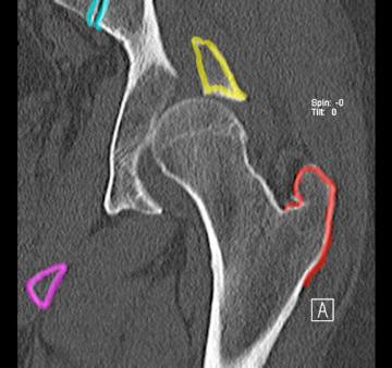
This is a coronal CT image on our patient, showing upward displacement of the femur, with the pubic bone outline in purple, sacroiliac joint in aqua, bone fragment in yellow and greater trochanter in red. CT is a useful way to image bony alignment and fractures, but is not as good as MR for imaging tendons, cartilage and ligaments. |
|||||||
B
A
CASE 1 followup