Tufts Anatomy Yr-1 |
||||||||||
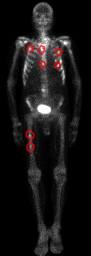
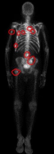
These images show diffuse spread of the patient's prostate cancer to many bones--metastases. The anterior view shows the lesions in the sternum and anterior ribs, as well as the right femoral diaphysis. The posterior view shows lesions in posterior ribs, pelvis and spine. The white area in the pelvis is the bladder. The tracer is eliminated by the kidneys so it ends up in the bladder. |
||||||
 |
 |
|||||
POSTERIOR view |
||||||||
ANTERIOR view |
||||||||
CASE 4 followup