PrISM Cardiovascular |
||||||||||||||
 |
 |
|||||||||
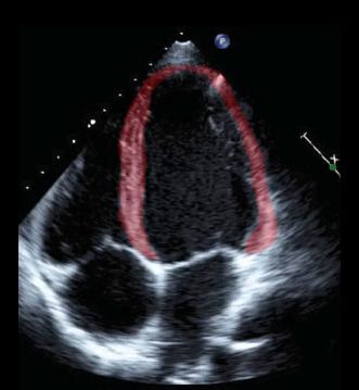
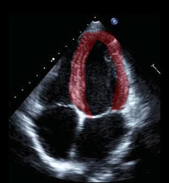
Image A shows ventricular diastole and Image B shows ventricular systole.The ejection fraction in this patient is over 50%. The range of normal is from about 50% to 75%. Is there any other imaging modality that can give you similar information about heart function? |
||||||||||
image A |
||||||||||
image B |
||||||||||