PrISM Cardiovascular |
||||||||||||||
 |
||||
 |
||||||||
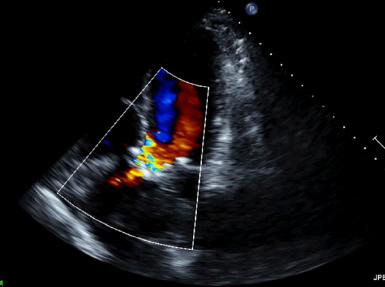
The top images show an aortic valve that barely moves, with a thin high velocity jet of blood shooting across the narrow opening. This patient would have a systolic murmur.
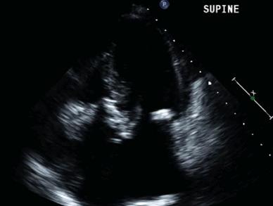
The bottom images show an abnormal mitral valve, with one leaflet that opens relatively normally, and another that is very thick and white (echogenic) with very little motion. This patient would have a diastolic murmur.