PA Anatomy: Imaging Overview-Nuc Med |
||||||
Nuclear medicine is also called 'functional imaging', as the images often reflect the function of some physiologic process within the body. Some tracers delineate normal anatomic structures, and some can be used to measure function, in a manner similar to ultrasound. Some tracers are taken up by abnormal processes, like tumors or infection. All nuclear medicine images are low resolution, and may only show one organ or structure, rather than the entire body. In order to interpret them, you need to know a lot about the tracer that was used. |
||||||||||
 |
||||||||||
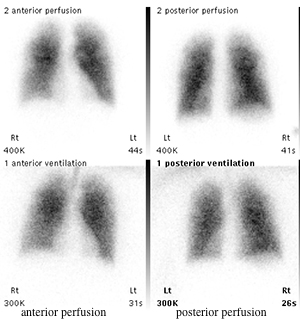
This study is called a 'ventilation-perfusion scan', or VQ scan. The top two images show where air goes within the lungs, and is obtained by having the patient breath in radioactive gas. The bottom two images use a tracer that shows where the blood is flowing in the lungs. The two sets of images should look identical, as they do in this normal study |
||||||||||
 |
||||||||||
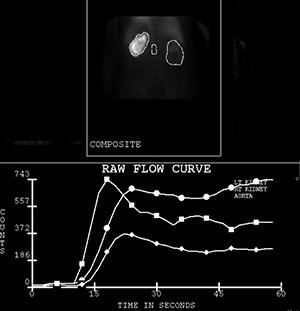
This is a renal scan, where the tracer is taken up by the kidneys and excreted, which can be quantified, as shown in the graph at the bottom. This study is NOT normal, as the right kidney shows quite a bit of uptake (white on the top image) but the left kidney shows much less. The kidneys are outlined on the top image to allow calculation of flow. |
||||||||||