PA Anatomy: Imaging Overview-Nuc Med |
||||||
 |
||||||
Nuclear medicine images are fundamentally different from other types of diagnostic imaging, because they use radioactive agents, and the radiation is coming out of the patient instead of being beamed through them from an external source. Special cameras are used that can precisely locate the source of radiation within the body to generate the images, but the images will look different if the camera is brought close to one side of the body vs the other, as the radiation will be partly absorbed and scattered by body tissues. The sharpest image will be when the camera is closest to the source of the radiation. |
||||||
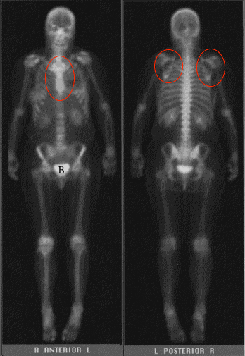
This is an example of a type of nuclear medicine imaging called a 'bone scan'. The radioactive tracer has been designed to home in on bone, and will be particularly evident in areas where bone is being remodeled, which happens normally near joints. It will also home in on areas where bone is being remodeled due to abnormal conditions, like tumors or infection. This is a normal study. Notice that there is a lot of activity or uptake in the lower pelvis, which is the bladder (B). The tracer is eliminated from the body by the kidneys, and excreted into the urine. Note that the sternum is best seen on the anterior view and the scapulae are best seen on the posterior view (circles). |
||||||