PA Anatomy: Imaging Overview- MR |
||||||
 |
||||||||
MR is different from CT in many ways, but one is that we do not use different 'windows' to look at images. For each different type of tissue we are interested in, we do a separate image aquisition, which means that a complete MR series can take up to an hour or more to finish. Two common image seqences that are used in MR are T1-weighted and T2-weighted images. To tell these apart, you need to find some water or watery fluid somewhere on the image. This could be around the spinal cord, in the gallbladder, in joints, or in the bladder, for example. |
||||||||
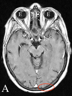
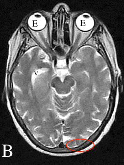
These are two sequences from a brain MR. Each was acquired separatelyl If you look at the eyeballs (E), which contain fluid, you can see that they look very different in Image A compared to Image B. Image A is a T1-weighted sequence and Image B is a T2-weighted sequence. There is also fluid that surrounds the brain, the cerebrospinal fluid. On Image A, a bit of this fluid is seen in the ventricle of the brain (V) and is low signal. In Image B, this same fluid is high signal (V). Notice the skull (oval circle) on both images. The outer and inner edges of the skull bone are dark (low signal), regardless of whether the image is T1 or T2-weighted. Fat is relativel high signal on both image sequences as well. You need to find FLUID to tell T1 from T2-weighted images. |
||||||||
 |
||||||||