PA Anatomy: Imaging Overview- MR |
||||||
CT and MR images can look similar at first, as they are both slices, and can be displayed in any of the anatomic planes already discussed (axial, sagittal, coronal). The best way to tell a CT from an MR is to look at bone, in particular the outer rim of bone, called 'cortical bone'. On CT, this is normally the whitest thing on the image. On MR, the outer edge of bones is very dark. Other tissues on MR may look similar to CT or very different, depending on how the study was done. For example, fat is often very white on MR (but not always), and is ALWAYS dark on CT. So bone is more reliable to look at. |
||||||||||||
 |
||||||||||||
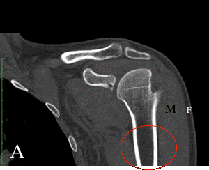
On CT, fat (F) is always darker than muscle (M) |
||||||||||||
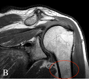
These are both coronal images of the left shoulder in different patients. Image A is a CT (bone window, since you can see lower attenuation in the center of every bone). Notice that the outer edges of all bones are white-the whitest thing on the image. Image B is an MR sliced at a slightly different angle. Notice the outer portion of the bone along the shaft of the humerus (inside the red oval). The outermost bone is very dark, and only the central part of the bone is white. It is the reverse of the CT pattern. |
||||||||||||
 |
||||||||||||
On MR, fat is often white, but on some sequences this is not true. In this image, fat (F) is very white, or 'high signal' |
||||||||||||