PA Anatomy: Imaging Overview- US |
||||||
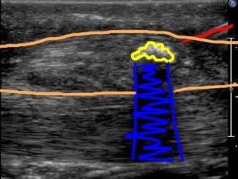
Ultrasound is a bit like fluoroscopy, in that during the actual exam, the operator sees what is going on in real-time, and captures selected images to show what they saw. This image series shows a needle being inserted into the left Achilles tendon, to break up some calcification that has formed there, and relieve pain. A labeled image is shown below the movie to help figure out what is going on. US images take practice to interpret because they are low resolution (not much detail). The best view of what is going on is obtained by the operator who sees everything in real-time, as it happens. |
||||||||
Because US is a real-time exam, it is ideal for guiding procedures, since you can watch where the needle throughout the entire procedure. |
||||||||
 |
||||||||
yellow=calcium deposit; blue=blocked beam beyond the calcium (which acts like a bone); red=needle; orange=Achilles tendon |
||||||||