PA Anatomy: Imaging Overview- US |
||||||
 |
|||||||||||||||||||
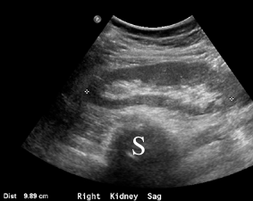
This is a sagittal image of the right kidney, with calipers on the image to measure the length of the organ from top to bottom. The spine (S) is the black area at the bottom of the image. The kidney has an outer dark grey part (called the cortex) surrounding a bright white, echogenic area (called the renal sinus). |
|||||||||||||||||||
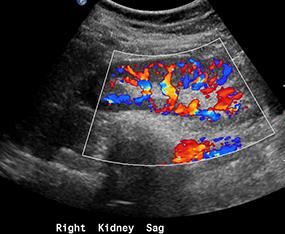
US can also be used to quantitate various bodily processes, such as blood flow. There are several ways to do this. One is to use 'color flow' imaging, which shows movement of fluids like blood either toward or away from the transducer using a scale from blue to red. Another way is to pick a small area of the image and display movement within that region as a graph. The ability to quantitate flow is what makes US so useful in imaging structures like the arteries in the neck and the heart. |
|||||||||||||||||||
 |
|||||||||||||||||||
This is the same kidney, with color flow imaging. You can see how dramatic the vascularity is within this organ. The color scale does not show arteries as red and veins as blue, but the color differences relate to whether the flow is toward or away from the transducer. |
|||||||||||||||||||
 |
|||||||||||||||||||
 |
|||||||||||||||||||
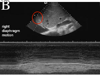
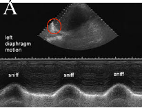
These two images are from a study called a 'sniff test'. This is done to document motion of the diaphragms. Image A shows the left diaphragm, which has the detector overlying it (oval). When the patient sniffs, the graph at the bottom shows normal motion. Image B shows no motion, or paralysis. |
|||||||||||||||||||