PA Anatomy: Imaging Overview- CT |
||||||
CT data shows the average density of body tissue within a tiny square, called a pixel. There is a wide range of densities that can be detected with CT, and if all are displayed, the image has a washed-out look. So we routinely set the brightness and contrast of CT images to optimize view of some of the types of tissue that we are most commonly interested in seeing. Each of these tissue-specific brightness/contrast settings is called a 'CT window'. They can be changed after the study is done and the patient has left, since they are using the same data, just displayed in a different way. |
|||||||||||||||||||
 |
|||||||||||||||||||
BONE window-notice that you can see a central marrow cavity that is lower attenuation than the outer cortex in every bone on the image. Notice that fat and muscle are similar shades of grey. |
|||||||||||||||||||
 |
|||||||||||||||||||
LUNG window-this shows all of the tiny bronchi and vessels in the lungs, as well as pleural lining, but is not good for looking at other types of tissue, like the chest wall (again, fat and muscle are similar shades of gray) |
|||||||||||||||||||
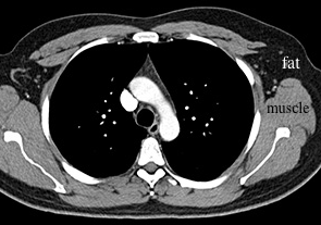
SOFT TISSUE window-does not show lungs very well, does not show marrow cavities in bones (but bones are still visible, just no details), but very good for showing muscles and organs, particularly when outlined by fat, because they are very different in darkness (fat is much darker than muscle) |
|||||||||||||||||||
 |
|||||||||||||||||||
All three of these images show the exact same data, with different brightness and contrast settings. There are other special CT windows for brain, liver and contrast material. |
|||||||||||||||||||