PA Anatomy: Imaging Overview- CT |
||||||
There are three standard anatomic planes that are also used to display data for CT scans: horizontal (or axial) coronal (cutting the body into slices that go from anterior to posterior or posterior to anterior) sagittal (cutting the body into slices that go from right to left or left to right) CT data can also be displayed in oblique or even curved planes, and can be reconstructed into 3D representations of surfaces |
||||||||||||||||||||||
 |
||||||||||||||||||||||
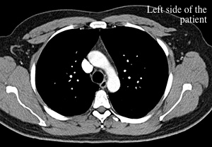
Axial CT slice through the upper chest. Notice that bones are very white. Just like for a CXR, the LEFT side of the patient is on the RIGHT side of the image. Imagine you are going in to examine a patient who is laying on an exam table. You are coming toward their feet, and the LEFT side of their body is to your RIGHT. It is just the same as the CT orientation |
||||||||||||||||||||||
 |
||||||||||||||||||||||
 |
||||||||||||||||||||||
 |
||||||||||||||||||||||
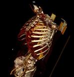
3D model of the skeleton from this CT data set |
||||||||||||||||||||||
Coronal slice through the region of the heart. Once again, the patient's LEFT is to the RIGHT on the image, as if you are facing them and talking to them. |
||||||||||||||||||||||
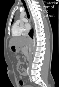
Sagittal slice through the region of the heart, with the patient facing to the left and their spine to the right side of the image. |
||||||||||||||||||||||