PA Anatomy: Brain: Case 2 |
||||||
 |
||||||||
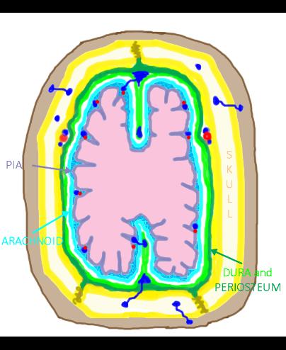
To figure out where the abnormal blood is located, we must review the meningeal linings of the brain, and their relationship to blood vessels. |
||||||||
The blood in this case is in the SUBDURAL space, a potential space between the dura (which is tightly adherent to the inner table of the skull) and the arachnoid, which covers the brain but does not extend down into the sulci. The typical shape of a subdural hemorrhage is a crescent shape. |
||||||||