PA Anatomy: Brain: Case 2 |
||||||
 |
 |
|||||
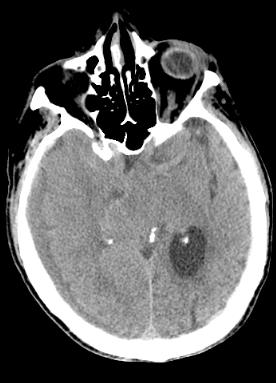
On a CT of the head without IV contrast (basilar and middle cerebral are only faintly seen), all abnormal white areas should either be acute bleeding or calcifications. Calcifications are whiter than acute blood, around 200 HU, similar to bone. |
||||||
The left lateral ventricle is large, but the right one is not apparent (compressed). The bright white areas on this scan are calcifications (circled in purple), in the choroid plexus of both lateral ventricles and in the pineal gland. |
||||
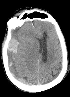
The lateral ventricles are also very asymmetric on this scan, and significantly shifted to the left, indicating increased pressure. There is old blood and new blood. |
||||
Where is this abnormal blood located??? |
||||||