Anatomy |
||||||||||||||||||||
 |
||
 |
||
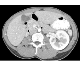
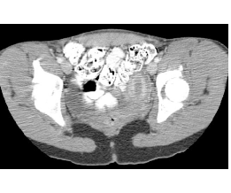
This patient was born with only one kidney on the left. Because the urinary and genital systems develop together, it is important to look for abnormalities of the uterus in patients with renal anomalies. In the pelvis in this patient, the uterus is an unusual shape, consistent with a unicornuate uterus, missing the right half. The IVC, left renal vein and external iliac veins, external iliac arteries and rectum are also shown on Image F, with unlabeled imag E.
Note the difference in appearance of the renal cortex and medulla on Images C and D. This is because the scan was done very soon after injection of contrast into an arm vein. The cortex lights up before the medulla, just as the aorta is higher attenuation than the IVC.