Anatomy |
||||||||||||||||||||


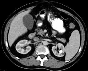
This is a CT with IV contrast and oral contrast, not ideal for identifying stones. If you want to detect tiny stones, CT is more sensitive than a KUB and the best way to do the CT scan is without oral or intravenous contrast, so that any white stone will stand out more. White oral or IV contrast can actually hide small stones.
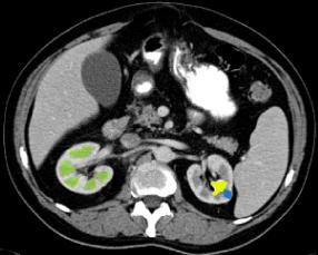
The study was done early after IV contrast injection because you can clearly tell the medullary parts of the kidneys, which show up a pyramid-shaped dark triangles (green on Image B). The stone (yellow on Image B) is clearly visible but there is no dilatation of the collecting system. There is a small incidental left renal cyst (blue on Image B). A stone as large as this one is unlikely to enter the ureter, but could cause an obstruction at the uretero-pelvic junction if it became dislodged from the renal tissues and moved to the region where the upper ureter joins the renal pelvis.
What other type of imaging could be done to assess for possible renal obstruction in patients suspected of having kidney stones?
A
B