Anatomy |
||||||||||||||||||||
 |
||||
 |
||||
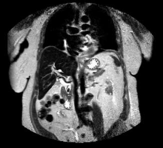
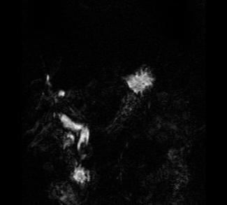
These are both MR images. The image on the left is a coronal T2-weighted sequence, with bright fat and high signal fluid in the stomach and biliary system. The image on the right is a 3D reconstruction that just shows the biliary system, an MRCP. Both studies show a dilated biliary tree and a low signal lesion in the distal CBD consistent with a stone. MR is usually not the first study done in patients with jaundice, but can be very useful in complex cases where US or CT do not give a clear diagnosis.