Anatomy |
||||||||||||||||||||
 |
||
 |
||||||||
 |
||||||||
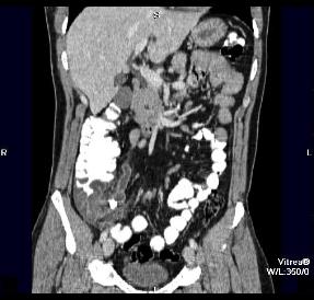
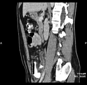
Our patient was NOT pregnant, so she had a CT with both oral and IV contrast (probably not optimal).
These are two representative images from her study. Decide what the imaging plane is for each and what is abnormal before bringing up the labels.
The cecal wall is shown in red, the appendix in green and an important muscle in orange. Why is this mucle important in terms of the patient's physical exam?
What is indicated in blue on the labeled images?