Anatomy |
||||||||||||||||||||
 |
||
 |
||
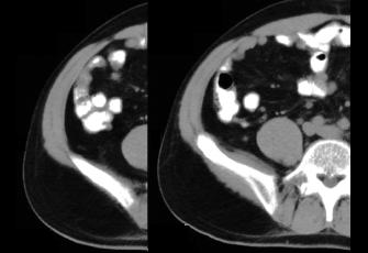
Both studies are normal. The top image shows a CT in the axial plane, soft tissue windows, with oral but without IV contrast. On the labeled image, the appendix is shown in green and is thin and sharply marginated (no stranding in the adjacent peritoneal fat to suggest inflammation). The terminal ileum is shown in yellow and the cecum in blue.
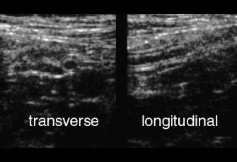
The bottom image shows the normal appearance of the multi-layered wall of the appendix on ultrasound images at the tip of the green arrows on the labeled image, in the planes transerve to the appendix and longitudinal to the appendix. It is hard to see the normal appendix on ultrasound, so ultrasound is better for demonstrating an abnormal appendix than for showing a normal one.
The MOST IMPORTANT question to ask this patient is whether she might be PREGNANT! We do not want to do a CT in that situation!!