|
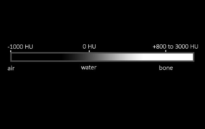
On CT, white matter is lower in density (darker) than grey matter because it consists of both nerve tissue and myelin, a fatty substance. Fat is lower in density than water (butter floats on water when mixing up batter for a cake). It is important to realize when looking CT imaging, that the density of flowing blood is different from clotted blood. On CT, acute hemorrhage is usually slightly higher density than normal blood in vessels, but over time, it will become closer to water density. IV contrast is much denser that blood or any other tissue (except calcifications).
|
|
