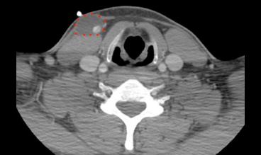
A-water (the lump was a cystic, fluid-filled lesion; there is a small area of calcification in the mass posteriorly as well)
B-fat (thin layer of subcutaneous fat with platysma running through it)
C-muscle (the sternocleidomastoid muscle on the left)
D-contrast in the internal jugular vein (not very dense, because not much contrast was given, but the vessel is definitely denser than adjacent muscle)
E-bone (cervical vertebral body)
F-metal (skin marker placed over the lump)
This patient is relatively thin (little fat) but has good muscle mass.
|


