|
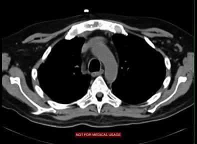
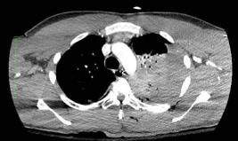
Recognizing CT images takes a bit of practice. The bones are usually the easiest thing to see on the image, which is a clue. Recognizing various windows also takes practice. If the muscles stand out against a dark background of low attenuation fat, then it is probably a soft tissue window. This type of image is not good for looking at bones or lungs, just soft tissues. The area outlined in blue is the metal button we saw on the previous image. This patient, as we suspected on the topogram, does not have very high muscle mass. The pectoral muscles are thin on both sides.
|
|


