ICM-II: CHEST, case 2 |
||
 |
||||
 |
||||
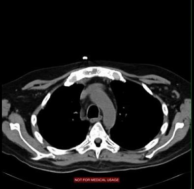
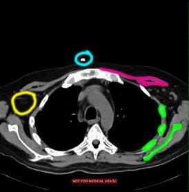
You can tell that this is a CT (not an MR) because the outer cortical bone is very high in attenuation (shown in green). The imaging PLANE is axial or horizontal. The WINDOW is a soft tissue window (because the apparent attenuation of muscle (pink) is very different from fat (within the yellow circle). There is no IV CONTRAST present because the aorta is the same attenuation as muscle. Can you tell on this image whether oral CONTRAST was given? What is outlined in blue? |
||
 |
|||||||||||