Set 5: hilar angle

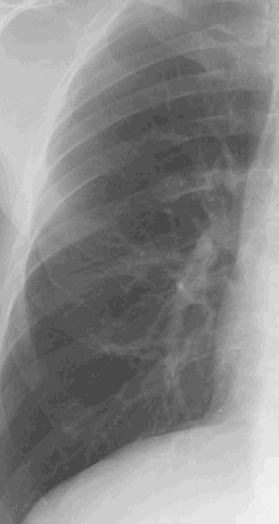
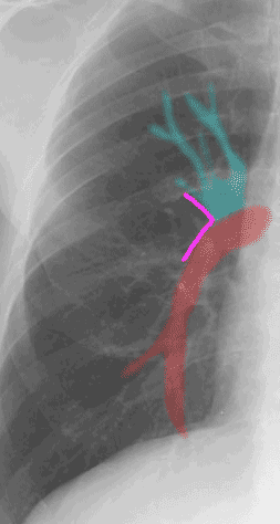
The hilar angle is an anatomic landmark that can help you to start making sense of one of the most confusing areas of the chest radiograph--the hilum. The hilum is very hard to assess because it varies a lot in terms of shape, position and size from one patient to the next. But most often there is a distinct point at which upper lobe vessels cross the large vessel going to the lower lobe, called the interlobar pulmonary artery, shown in red. Looking for the hilar angle can help you decide if vessel margins are sharp, and can remind you to compare upper lobe vessel diameter (above the hilar angle) to lower lobe vessel diameters when assessing for possible CHF.
