Set 5: vascular distribution

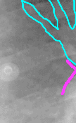
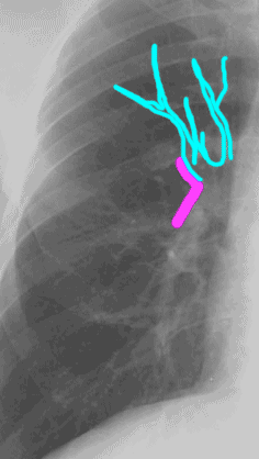
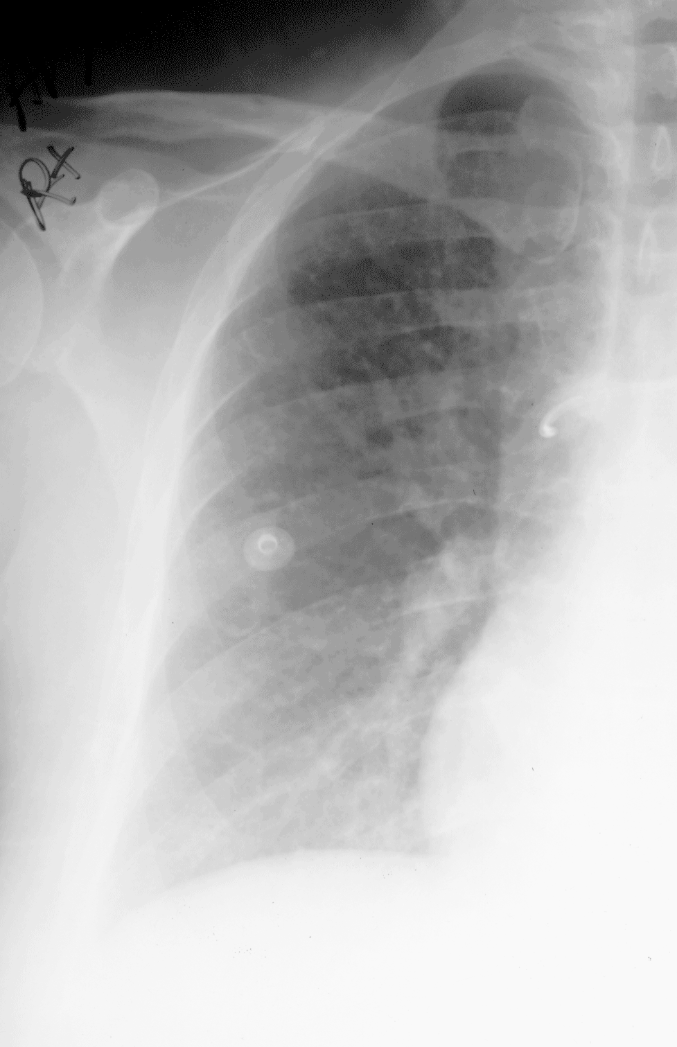
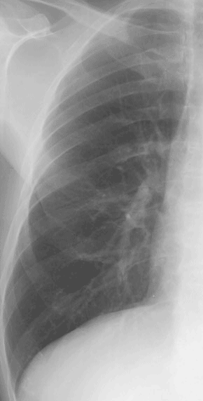
In these two examples, upper lobe pulmonary vessels are outlined in blue. They are much larger in Image A than in Image B, and overall, vessels are fuzzier and harder to see the outlines of in Image A than in Image B. This happens in CHF as the pressure builds up in the pulmonary circuit, causing upper lobe vessels to enlarge and fluid to leak out into surrounding tissues, making vessel margins fuzzy. The purple outline indicates an anatomic landmark called the HILAR ANGLE.

A
B

