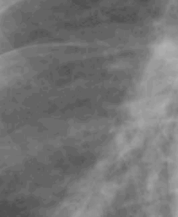
Set 2: interstitial edema

This set of images explores why the lung looks the way it does when something causes thickening of the interstitium, most commonly fluid from CHF. The most specific finding that tells you that a lung abnormality is INTERSTITIAL in location is the presence of Kerley B lines. See if you can find them in this example.


