Set 2: CT of interstitium

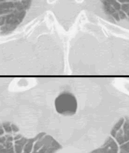
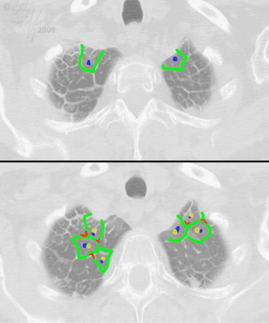
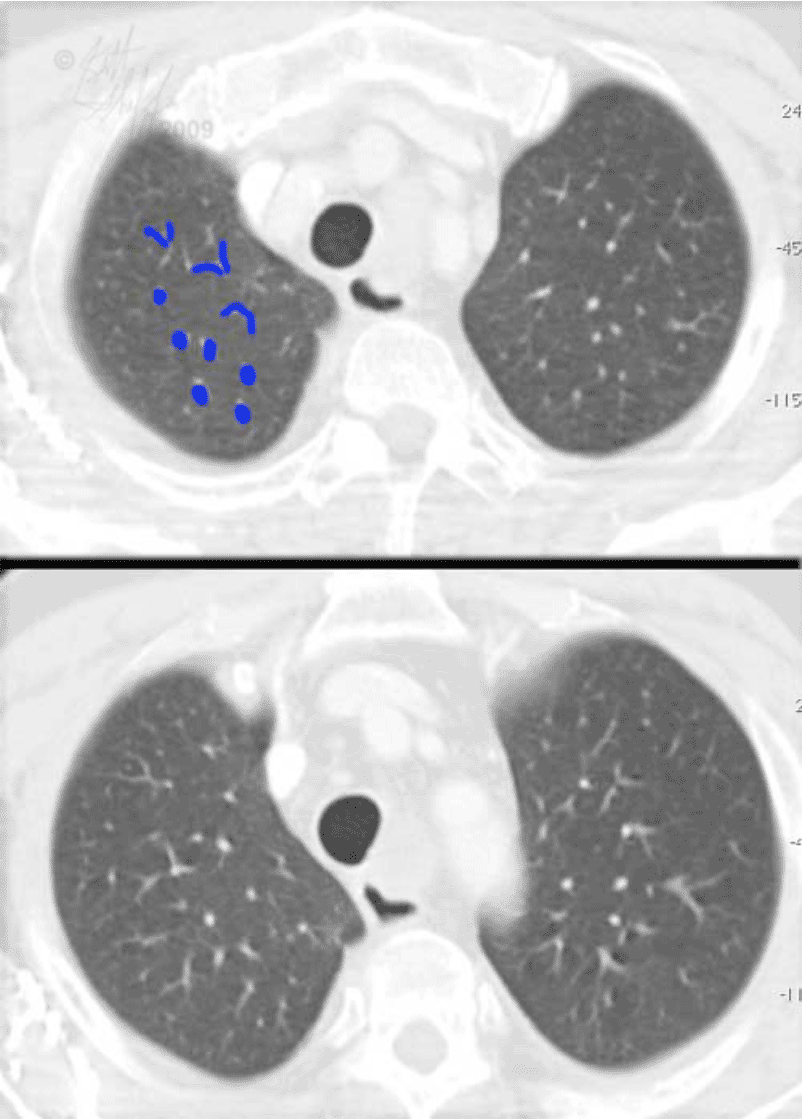
Kerley B lines are a radiographic finding, but thickening of interlobular septae can be seen even better on CT, as shown in this example. See if you can identify the septae and the pulmonary artery branches on these two images. In this case, the thickened interlobular septae were due to spread of tumor to the lung, bronchoalveolar cell carcinoma, and were not related to CHF. As previously mentioned, interstitial patterns in the lungs can be due to many causes.

