Set 2: CT of interstitium

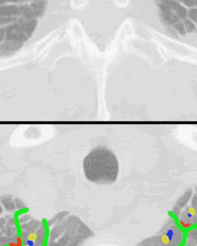
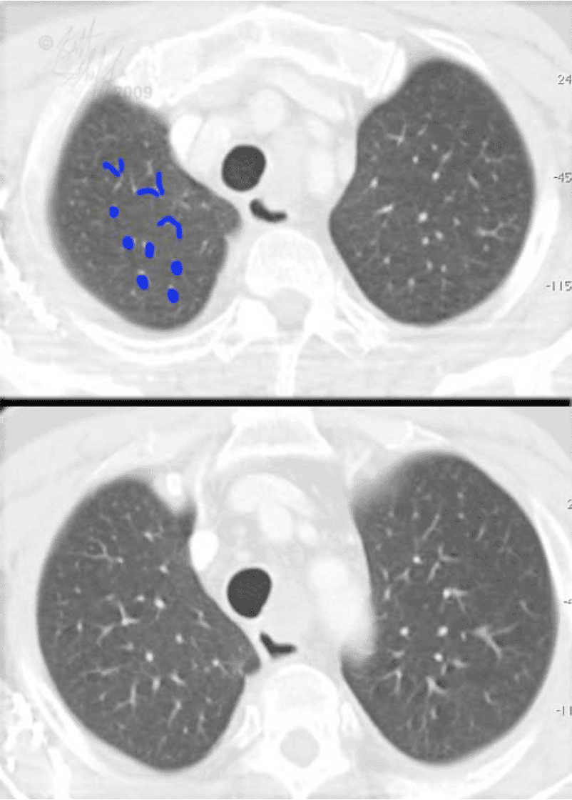
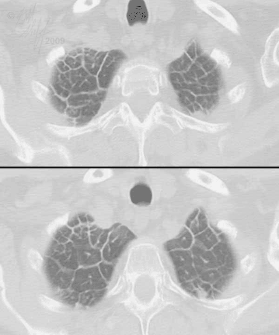
The interlobular septae form characteristic polygon-shaped structures when thickened on CT, best seen at the edges of the lungs. The thickened septae are shown in green, the central pulmonary artery branch in blue, the bronchus in yellow, and the location of pulmonary veins in red. The veins cannot be distinguished from the septae in which they run, and the bronchi are too small to be visible. This diagram shows you where they are located, even though you cannot see them. In normal lung, only the pulmonary artery branches are visible in the periphery of the lung. Tiny bronchi, veins and interlobular septae are too small to be visible normally.

