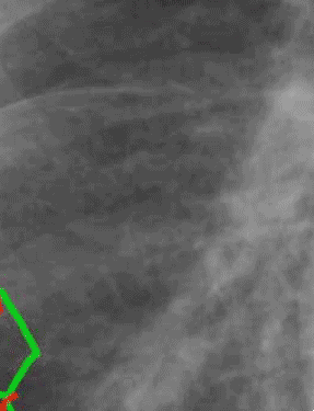
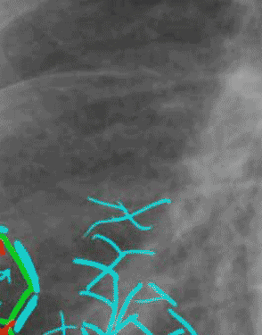
Set 2: interstitial thickening

In these diagrams, two secondary pulmonary lobules have been shown in enlarged diagrammatic fashion to highlight their internal structure. In the center of each lobule is a pulmonary artery branch (shown in dark blue), with an accompanying small bronchus (shown in yellow). The pulmonary veins (shown in red) run in the connective tissue struts (shown in green). In normal patients, none of these structures are visible at all in the outer lung on radiography. On the right is demonstrated what happens when a patient has chronic or subacute pulmonary edema. Fluid leaks out of the vascular system (light blue arrows) and makes its way to the interstitium, causing thickening. This is visible on the chest radiograph as Kerley B lines in some cases. These are best seen in the costophrenic regions because of their orientation. In other parts of the lung, the thickened interlobular septae are more randomly oriented, and contribute to the appearance of a reticular pattern, as shown in light blue on the right image.
