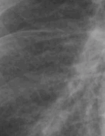
Set 2: interstitial edema
secondary pulmonary lobules

Here is our abnormal case again, with the visceral pleural surface shown in yellow. The green outlines represent the connective tissue struts of the basic functional unit of the lung, called the ‘secondary pulmonary lobule’. These are MACROscopic structures, about a centimeter in diameter, as shown here. In normal patients, they are not visible. They can become visible due to edema in patients with CHF, that tracks along these connective tissue pathways, as in this case. Because of their orientation perpendicular to the pleural surface, they are easiest to see in the lower lateral lungs (but they are present all over).
