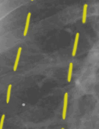
Set 2: interstitial edema
lung zones

In this normal case, it is much easier to see the margins of the large vessels in the inner zone, and to trace out the branches in the middle zone. There are no markings in the outer zone. Compare this to the abnormal image. Notice how much clearer the vessel margins are in the inner and middle zones in this normal example.
