|
The labeled images from the lower abdominal CT show that the anterior abdominal wall is weakened where the rectus abdominus was removed (red dotted line) and the other abdominal muscles (outlined in red) are inadequate to keep the viscera in place. Many bowel loops have herniated out (blue arrows). This is a known complication that can occur after TRAM surgery, and some surgeons prefer to do a more complex free-flap procedure (DIEP or deep inferior epigastric perforator) where a perforating artery is dissected out and cut loose from the muscle, taking the overlying fat along. This can then be connected to vessels in the breast region using microsurgical techniques.
|
|
|
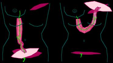
This diagram shows the vascular supply (in green) to rectus abdominus, from both ABOVE (via the internal thoracic artery, feeding into the superior epigastric artery) and from BELOW (via the inferior epigastric artery). So if the muscle is cut from its inferior attachment, it can still retain blood supply from above.
|
|
|


