Anatomy Yr-1 |
||||||||||||||


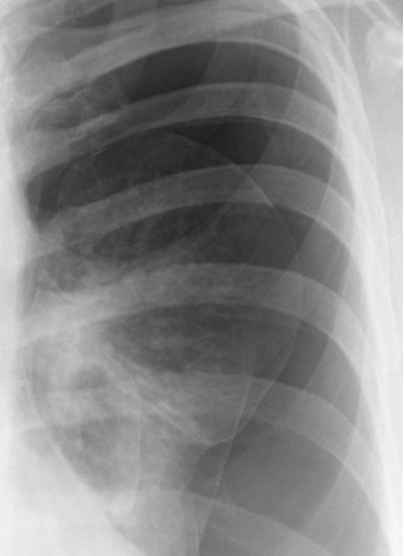
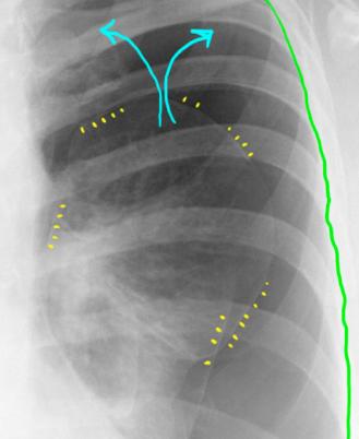
Image A is a closeup view of the left lung in our patient. If you look at the edge of the lung, you can see a thin white line (indicated by the yellow marks on Image B). This represents the visceral pleura and underlying connective tissue. The parietal pleura (green) remains attached to the inside of the ribs.
The air (blue arrows) is collecting between the visceral and parietal pleura, from a tear in the visceral pleura which is too small to be visible. Without the normal vacuum in the pleural space, the elastic recoil of the lung tissue causes it to contract down and collapse. In most cases of pneumothorax, the air in the pleural space escapes from the LUNG, and is not introduced from outside. Any track to the outside usually closes up right away.
A
B