Anatomy Yr-1 |
||||||||||||||


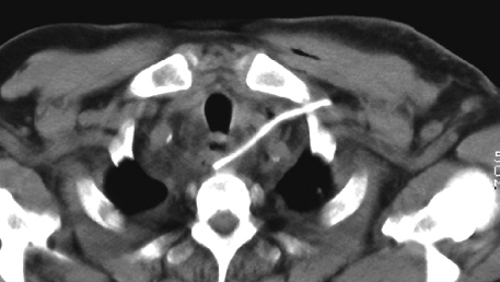
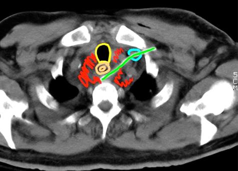
Image A is an unlabeled axial CT without IV contrast, soft tissue windows. Image B shows labels:
yellow-trachea
orange-esophagus
green-catheter
blue-left brachiocephalic vein
red-hematoma
This catheter started out in the subclavian vein on the left, but then exited the vein and passed through the mediastinum to end with its tip posterior to the esophagus, producing a lot of bleeding in the region. It was removed and the patient recovered.
A
B