Anatomy Yr-1 |
||||||||||||||

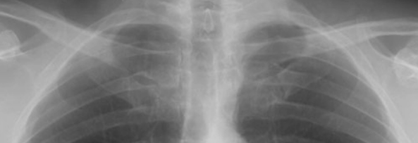
Comparing Image A of our patient's lung apices to Image B of a normal patient, you can see that the top of the air-filled lung on the left is LOWER than on the right in Image A. The difference is subtle but important. This indicates blood in the extrapleural space, probably coming from the mediastinum or neck.

A
B